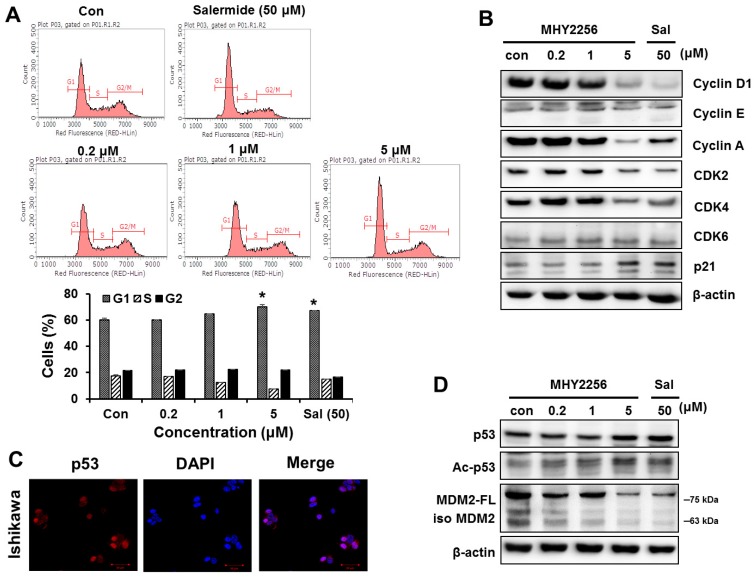
Figure 2.
MHY2256 increases G1 arrest and reduces p53 levels via mouse double minute 2 (MDM2) degradation. (A) The Ishikawa cells were treated with the indicated concentrations for 48 h. The cells stained with propidium iodide (PI) were subjected to flow cytometric analysis in order to determine their distributions in each phase of the cell cycle. * p < 0.05 indicate significant differences between the control and treatment groups. (B) The effect of MHY2256 on the expression levels of cell cycle regulatory proteins. The cells were treated with MHY2256 (0.2, 1, or 5 μM) or salermide (50 μM) for 48 h, and then, protein levels were detected by Western blot analysis. Aliquots of proteins were immunoblotted with specific primary antibodies against cyclin D1, cyclin E, cyclin A, CDK2, CDK4, CDK6, and p21. (C) Basal expression levels of p53 protein in Ishigawa cancer cells. Immunofluorescence and fluorescence detection of p53 using rhodamine red-tagged secondary antibody were done using confocal microscopy (magnification ×400). (D) The effects of MHY2256 on expression of p53, acetylated p53 (Ac-p53), full length of MDM2 (MDM2-FL), and MDM2 isomers (Iso MDM2). The Ishikawa cells were treated with MHY2256 and salermide for 48 h, and then Western blot analysis was performed.

