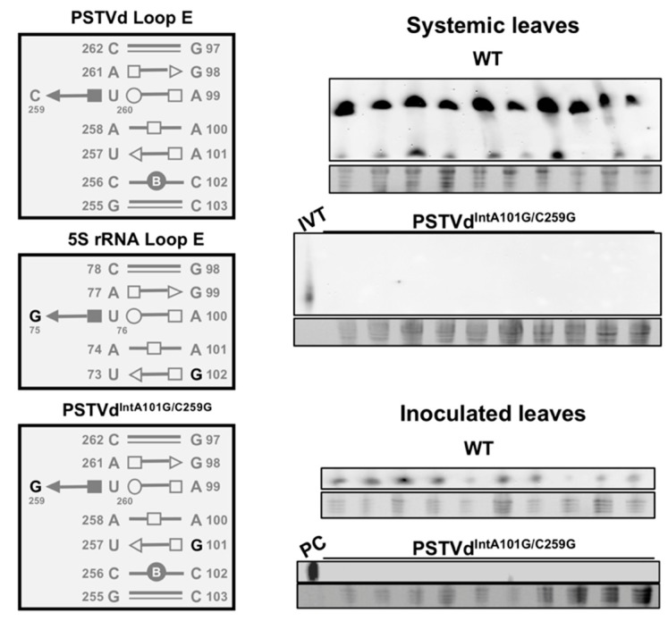
Figure 4.
Loss of infectivity of a potato spindle tuber viroid (PSTVd) variant with 5S rRNA loop E sequences. Left panel displays loop E structural arrangements in wildtype (WT) PSTVd, N. benthamiana 5S rRNA, and a loop E swapped PSTVdIntA101G/C259G. The right panel shows northern blots detecting the infection of WT PSTVd and PSTVdIntA101G/C259G in local and systemic leaves of N. benthamiana plants. PC, a verified RNA sample from PSTVd-infected leaves served as a positive control. IVT, the unit-length in vitro transcript served as a control. Note: only circular PSTVd (an indicator of replication) is shown in the northern blots for inoculated leaves. The “○, ⎕, ∆” symbols depict Watson-Crick, Hoogsteen, and sugar edges, respectively. The hollow and solid symbols represent trans and cis glycosidic orientations, respectively. These symbolic annotations of loop E 3-dimensional structures were previously explained in detail [63,65].

