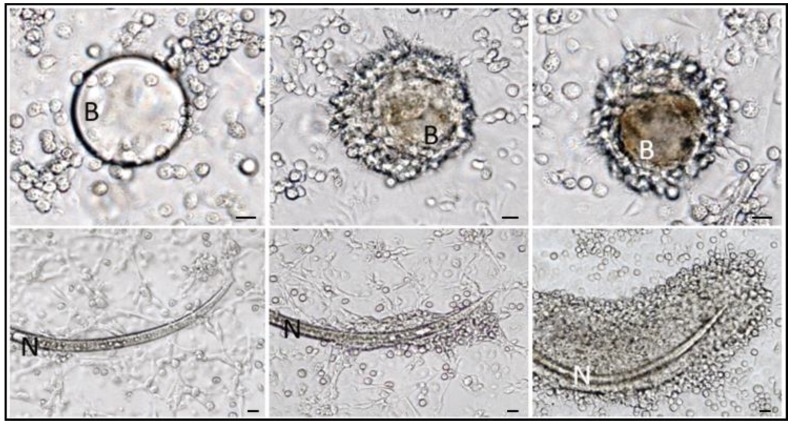
Figure 11.
G. mellonella hemocytes forming capsule around abiotic materials and free-living nematodes. (Upper) panels show encapsulation steps from 1 to 8 h after incubation of cultured hemocytes with synthetic beads (B); in the right micrograph is evident the melanin deposition around the bead inside the inner cell layer. In (lower) panels the same experiment was carried out with free-living nematodes P. rigidus (N). In both assays the progressive formation of multi-layered cellular capsules is observable. Bars = 50 μm (from [121]).

