Figure 3.
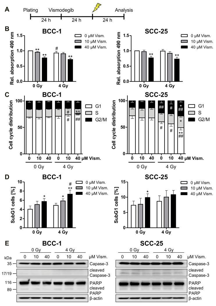
Vismodegib and irradiation modulate cell viability, cell cycle distribution and SubG1 cell fraction content. BCC-1 and SCC-25 cells were pretreated for 24 h with indicated concentrations of vismodegib or DMSO as control before a 4 Gy irradiation (A). At 24 h after irradiation, proliferation/viability was measured with a CellTiter 96® Aqueous One Solution Cell Proliferation (MTS) Assay (B). Cell cycle distribution (C) and SubG1 cell fraction (D) were analyzed after propidium iodide staining by flow cytometric quantification. Caspase 3 and PARP expression/cleavage was detected by Western blotting (n = 2) with β-actin as loading control (E). Data given in (B–D) are shown as means + SD from four independent experiments with quadruplicates (MTS assay, (A)) or duplicates (flow cytometry (B,C)). Differences were considered as statistically significant when * p < 0.05 or highly significant when ** p < 0.01; vismodegib- versus DMSO-treated cells (t-test). Significant differences between irradiated and non-irradiated cells are indicated as follows: # p < 0.05, ## p < 0.01 (t-test). Gy, Gray; PARP, poly ((adenosine diphosphate)ADP-ribose) polymerase; Rel., relative; Vism., vismodegib.

