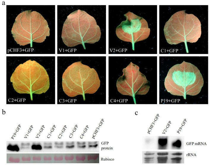
Figure 3.
Suppression of local PTGS by AGV V2. (a) The leaves of GFP-transgenic 16c line were co-infiltrated with agrobacterium suspension harboring 35S-GFP expressing GFP and one of the recombinant vectors expressing AGV proteins as indicated below the images. The leaves expressing pCHF3 + GFP were used as negative control and leaves expressing P19 + GFP were used as positive control. The photographs were taken under UV light at 5 d post infiltration. (b) Western blot analysis of GFP accumulation in the co-infiltrated leaf patches at 5 d post infiltration. The Ponceau-stained rubisco indicates the equal loading of total proteins. (c) Northern blotting analysis of GFP mRNA accumulation from the co-infiltrated leaf patches at 5 d post infiltration. The rRNAs below indicate the equal loading of total RNAs.

