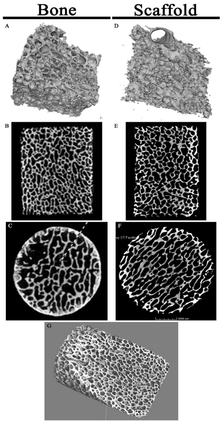
Figure 3.
Micro-CT Imaging of Specimens: Porcine cancellous bone micrographs (A–C) and scaffold micrographs (D–F) were similar in appearance on 3-dimensional projections (A,D), coronal (B,E), and axial (C,F) cuts. (G) High resolution scaffold 3-dimensional projection through the matrix interior displays the highly porous architecture.

