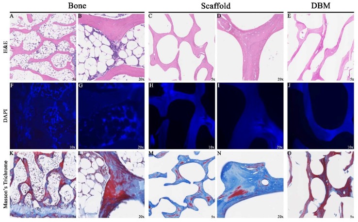
Figure 4.
Assessment of Decellularization by Histology: Representative sections 5 μm thick taken from specimen midsections showed removal of cellular contents (A,B) in the scaffolds (C,D). 4′,6-diamidino-2-phenylindol (DAPI) sections stained abundant cell specific material (F,G) which was not seen in scaffolds (H,I). Collagen content distribution was highly variable in specimens (K–N). Demineralized Bone Matrix (DBM) sections (E,J,O) were similar in appearance to the scaffold.

