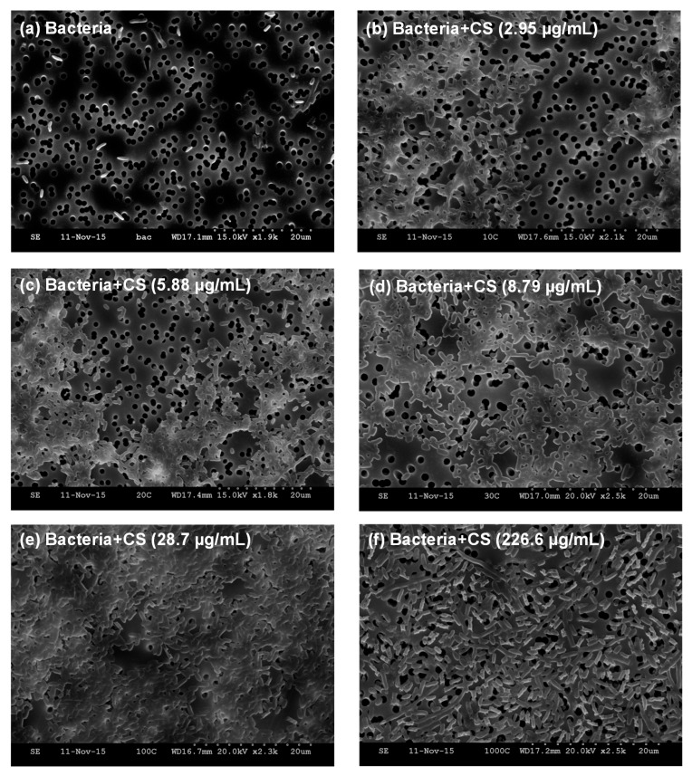
Figure 3.
Scanning electron microscopy images of (a) E. coli TOP10 bacteria alone; and an equivalent number of bacteria (OD600 = 0.2) mixed with varying concentrations of MDP DA30 chitosan, namely (b) 2.95 µg/mL; (c) 5.88 µg/mL; (d) 8.79 µg/mL; (e) 28.7 µg/mL; (f) 226.6 µg/mL, after incubation at 4 °C and slight shaking for 1 h, supported on track-etched polycarbonate membranes. Under these imaging conditions, bacteria appear as bright rod objects. Polycarbonate support appears as a gray background, and sharply-defined track-etched pores appear as dark circles of ~1.2 µm in diameter.

