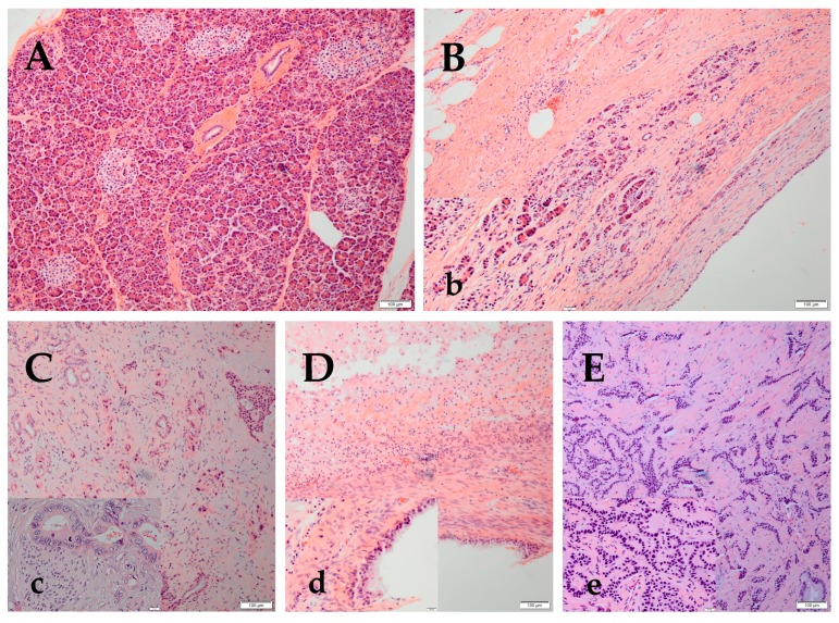
Figure 1.
Representative pictures from hematoxylin and eosin staining of normal pancreatic parenchyma, chronic pancreatitis (CP), pancreatic adenocarcinoma (PA), mucinous pancreatic cyst and pancreatic neuroendocrine tumors (NET). (A) Normal pancreas shows acini and interlobular ducts (exocrine) and islets of Langerhans (endocrine) (10×) (B) CP shows loss of acini and ductal tissue, as well as periductal fibrosis (10×). (b) The thumb image is a lower magnification of CP, depicting residual islets and interlobular ducts with flattened epithelium (40×). (C) PA is composed of small glands and malignant cell clusters with hyperchromatic nuclei invading in a desmoplastic stroma (10×). (c) The high magnification (40×) of a moderately differentiated PA shows glands composed of tall columnar cells with abundant cytoplasm. Perineural invasion, one of the characteristics of PA, is also seen here. The tumoral nuclei are large, with irregular nuclear membrane, frequently vesiculated chromatin, with numerous chromocenters and occasional proeminent, cherry red nucleoli. (D) The pancreatic mucinous cyst is composed of cells which contain intracytoplasmic mucin and fibrosed stroma (10×). (d) The high magnification (40×) shows mucin secreting glandular cells lining a benign mucinous cyst of pancreas (40×). (E) NET is composed of cells forming trabeculae, cords and ribbons of neoplastic cells (10×). (e) The high magnification (40×) photo shows a well differentiated NET of the pancreas; the cells are small to medium in size, with eosinophilic to amphiphilic and finely granular cytoplasm. The nuclei are monotonous, uniform, eccentrically located, round-to-oval, with “salt and pepper” (finely stippled) chromatin and no conspicuous nucleoli.

