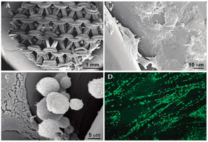
Figure 1.
Chondrocyte attachment on the knitted poly-l,d-lactic acid scaffold. (A) A knitted scaffold used for the cellular embedding. (B) A high proportion of the seeded chondrocytes (72%) adhered on the surface of the scaffold fibers within 12 h of cellular seeding, but most of the cells spread and flattened on the material after the initial adhesion. (C) Some chondrocytes could still adopt a spherical morphology. (D) Live/dead staining showed a good viability of the chondrocytes (green cells); however, the cellular attachment mainly occurred on scaffold fibrils, leaving most of the space unoccupied by the cells. Some red-stained dead cells were visible.

