Figure 2.
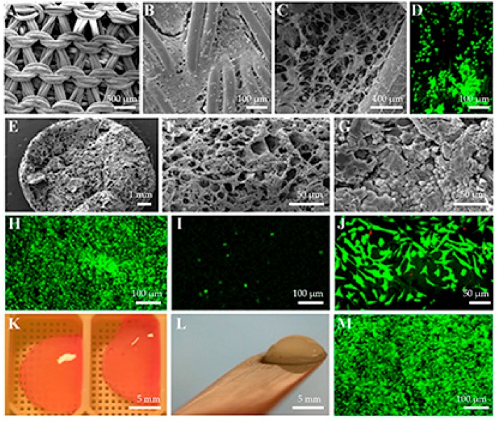
Examples of the use of recombinant human type II collagen as a biomaterial for primary chondrocytes. (A) The nonseeded knitted poly-l,d-lactic acid disc. (B) The surface of knitted poly-l,d-lactic acid disc filled with cross-linked recombinant human type II collagen seeded with primary chondrocytes. (C) The inner part of knitted poly-l,d-lactic acid disc filled with cross-linked recombinant human type II collagen seeded with primary chondrocytes. (D) The inner part of knitted poly-l,d-lactic acid disc filled with cross-linked recombinant human type II collagen seeded with primary chondrocytes and cultured with live/dead fluorochromes to visualize the live (green) and dead cells (red). (E) Recombinant human type II collagen sponge shows the porous structure of the material. (F) The porosity of recombinant human type II collagen sponge is also obvious inside the material. (G) The chondrocytes adhere well to the surface of recombinant human type II collagen sponge. (H) Live/dead staining indicates good cell viability on the surface of recombinant human type II collagen sponge. (I) However, only a few chondrocytes can reach the most inner part of recombinant human type II collagen sponge. (J) The chondrocytes seeded on the surface of recombinant human type II collagen membrane adhere well but show fibroblastic shapes and apparently dedifferentiated phenotype. (K) The chondrocytes mixed with soluble recombinant human type II collagen form the cell-embedded gels. (L) The chondrocyte seeded recombinant human type II collagen gels also stiffen during a two-week culture period. (M) The chondrocytes embedded in recombinant human type II collagen also have good cell viability for at least four weeks, as shown by live/dead staining.

