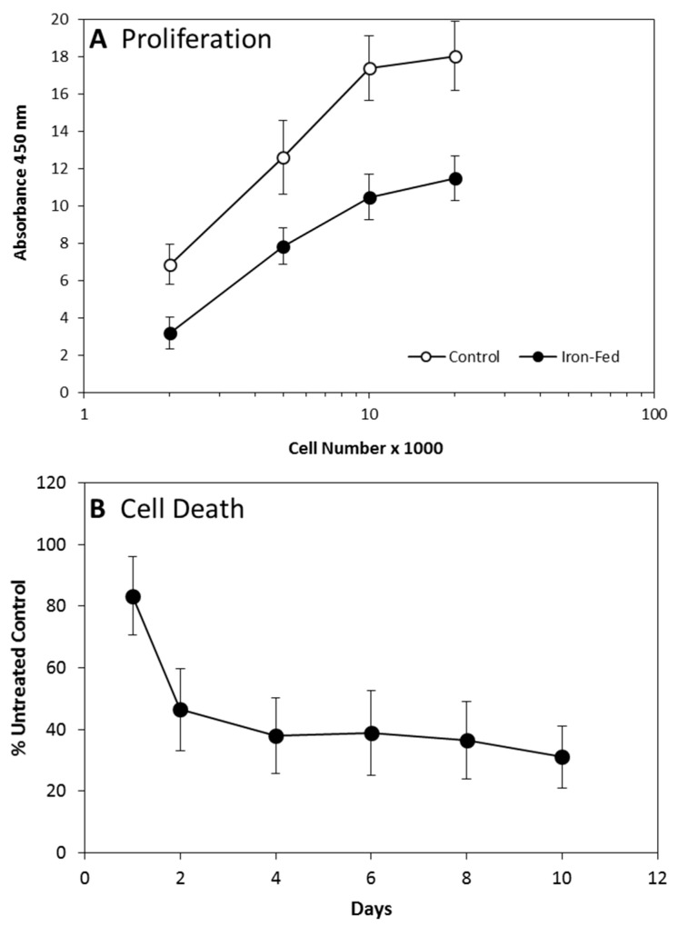
Figure 3.
Proliferation and Cell Death (A) The rate of proliferation of C8B4 microglia was assessed using a bromodeoxyuridine (BrdU) based ELISA kit. Both control and iron-fed microglia were plated into a 96 well plate at a range of densities and grown overnight. BrdU was then added for a further 16 h before the ELISA assay was used to assess incorporation levels. The level of incorporation was assessed by a colorimetric assay with a read out at 450 nm. Iron-fed microglia showed significantly lower levels of proliferation at all plating densities other than the highest (p < 0.05). Shown are the mean and S.E.M. of four separate experiments. (B) The level of cell death in C8B4 cells was assessed during their initial treatment with 500 μM ferric ammonium citrate. C8B4 cells were plated in 24-well trays at low density (20% confluency). The cells were then treated with iron for up to 10 days. The level of cell death was assessed using a commercial ELISA kit that determines the levels of histone associated DNA fragments in the cytoplasmic fraction. The results showed a significantly lower level of cell death in cells treated with iron for 2–10 days (p < 0.05). Only cells treated for one day showed no significant difference to the untreated cells (p > 0.05). Shown are the mean and S.E.M. of four separate experiments.

