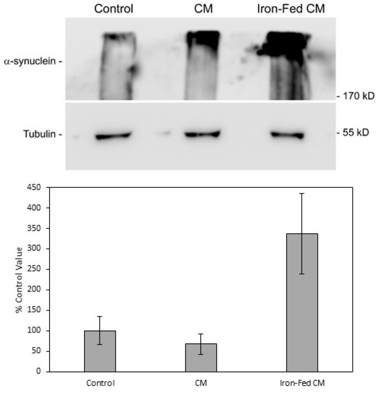
Figure 12.
Aggregation of α-syn. Western blot was used to detect high molecular weight aggregates of α-syn in extracts from SH-SY5Y cells treated with conditioned medium from primary microglia. SH-SY5Y cells were treated for 24 h with medium from control microglia (CM) or iron-fed microglia (Iron-Fed CM). Some cells were treated with just serum free medium (control). Extracts were prepared and electrophoresed on a 6% PAGE gel. The protein was then transferred by blot to a PVDF membrane (3 h, 100 mA) and α-syn detected with a specific antibody. High molecular weight bands for α-syn were indicative of aggregates. We also verified protein loading by re-probing the same blots for tubulin. Bands for α-syn were then analysed densitometrically. Values for control were normalised to 100% and values for the treated samples compared. Only treatment with iron-fed conditioned medium caused a significant (p < 0.05) increase in α-syn detected in the aggregate band. Shown are the mean and S.E.M. for four separate experiments.

