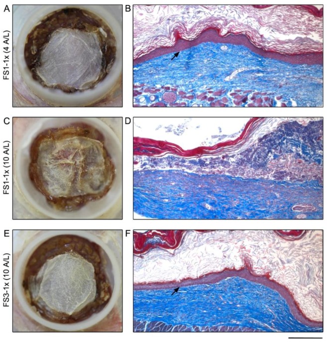
Figure 2.
Macroscopic and histological analysis of tissue-engineered skin substitutes matured 21 days in vivo. Representative macroscopic (left panel) and histological (right panel) results of FS1-1× (A–D) and FS3-1× (E,F) cultured for 4 (4 A/L) and 10 (10 A/L) days at the air–liquid interface before grafting in athymic mice. Arrows point out the fully differentiated epidermis well attached to the underlying dermis. Note that when the air–liquid interface culture period of FS1-1× was prolonged to 10 days before grafting, epidermis was absent in several areas after in vivo maturation (D). Histological coloration: Masson’s trichrome. Scale bar: A,C,D: 10 mm; B,D,F 235 μm.

