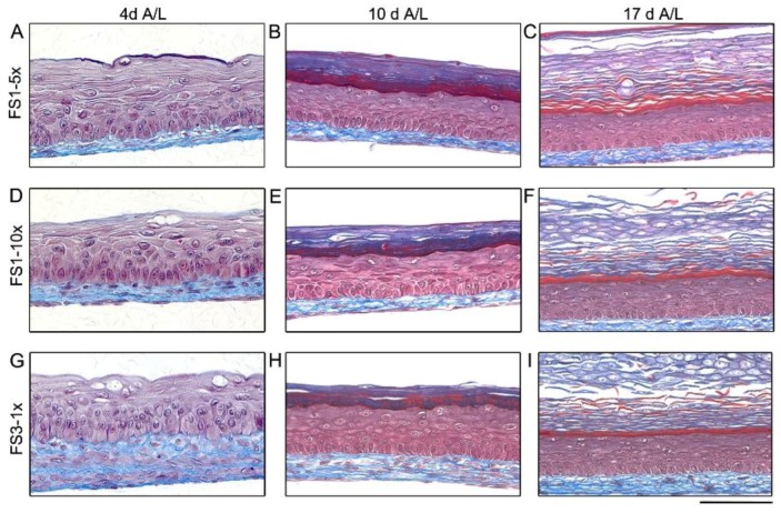
Figure 4.
Histological analysis of tissue-engineered skin substitutes matured in vitro. Representative histological results of TES produced with FS1-5× (A–C), FS1-10× (D–F), and FS3-1× (G–I) cultured for 4 (4 A/L), 10 (10 A/L) and 17 (17 A/L) days at the air–liquid interface. Histological coloration: Masson’s trichrome. Scale bar: 100 µm.

