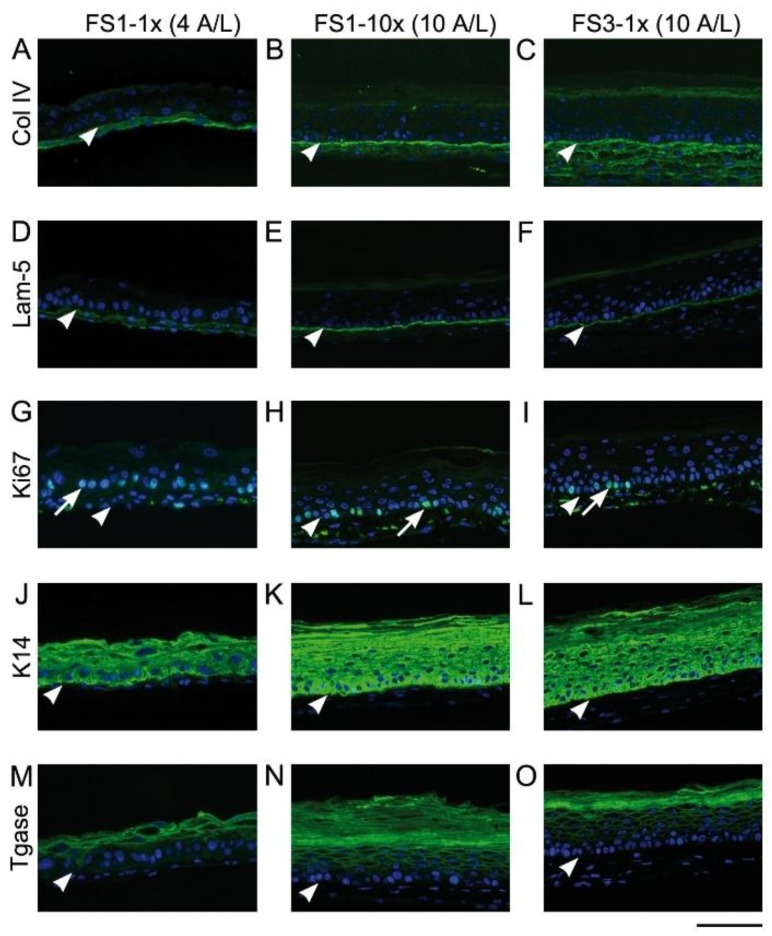
Figure 5.
Analysis of skin marker expression in tissue-engineered skin substitutes matured in vitro. Representative pictures FS1-1× (left panel), FS1-10× (center panel), and FS3-1× (right panel) cultured for 4 (4 A/L) and 10 (10 A/L) days at the air–liquid interface immunolabeled for the detection of type IV collagen (A–C), laminin 5 (D–F), Ki67 (G–I), keratin 14 (J–L), and transglutaminase (M–O). Arrowheads point out the level of the dermo-epidermal junction. Arrows point out nuclei expressing the proliferation marker Ki67. Col IV, type IV collagen; K14, keratin 14; Lam-5, laminin 5; Tgase, transglutaminase. Scale bar: 100 µm.

