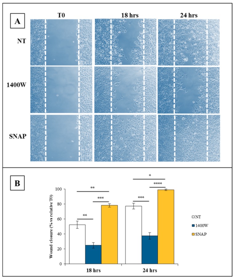
Figure 4.
Involvement of NO on the scratch-wound healing ability of U-87 MG cells. (A) Representative microscopy images of the scratch-wound healing assay captured at 0 h, 18 h, and 24 h. Scratched U-87 MG monolayers were incubated without (not treated, NT) or with NOS2 inhibitor 1400W (100 µM) or NO-donor SNAP (100 µM) for the indicated times after injury (10× magnification). (B) The extent of the wound closure rate was calculated as described and expressed as % closure versus relative T0 at 18 h and 24 h. Data are expressed as the mean ± SEM of two independent experiments in duplicate. For a comparative analysis of groups of data, repeated measures two-way ANOVA followed by a Bonferroni post hoc test was used (* p < 0.05, ** p < 0.01, *** p < 0.001, **** p < 0.0001).

