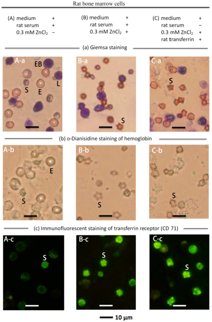
Figure 4.
Microscopic observation of cultured rat bone marrow cells with ZnCl2 supplementation. (A-a) Giemsa staining of the rat bone marrow cells (with medium and rat serum) without supplementation of ZnCl2 suspension-cultured for one day. The major cells were mature erythrocytes with a median cell diameter of 7.4 μm (E), lymphocytes with a median cell diameter of 7.3 μm (L), erythroblasts (EB), and smaller ~5 μm cells (S); (A-b) o-Dianisidine staining (to stain erythroid cells) indicated that mature erythrocytes (E) and the ~5-μm cells (S) were stained; (A-c) In CD-71 immunofluorescent staining, only the ~5 μm cells (S) were stained; (B-a) Giemsa staining of the rat bone marrow cells (with medium and rat serum) supplemented with ZnCl2 and suspension-cultured for one day indicated that most of the original mature erythrocytes (E) disappeared but many ~5 μm cells (S) appeared, and the lymphocytes (L) remained unchanged; (B-b) o-Dianisidine staining indicated that the ~5 μm cells (S) appeared multilobular and stained positively for hemoglobin; (B-c) The ~5 μm cells (S) showed strong immunofluorescence when stained with the CD-71 antibody; (C-a) Giemsa staining of the rat bone marrow cells in serum-free medium supplemented with ZnCl2 and rat transferrin suspension-cultured for one day. The cell pattern in (C-a) was similar to that of (B-a), i.e., many ~5 μm cells (S) appeared, and most of the original mature erythrocytes (E) disappeared. (C-b) o-Dianisidine staining indicated that the ~5 μm cells (S) appeared to be multilobular and stained positively for hemoglobin like those in (B-b); (C-c) The ~5 μm cells (S) showed strong immunofluorescence when stained with a CD-71 antibody.

