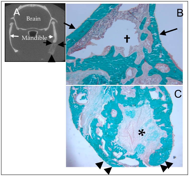
Figure 11.
Histological evaluation of the BMP-2-doped NBS from Figure 10. The non-demineralized sections are embedded in acrylic and stained with Goldner’s trichrome. Mineralized tissue stains blue. (A) This coronal μCT section provides orientation for B & C. The black arrows point to the NBS/bone interface (corresponding to those in (B)) with the implant seen beneath the arrows. The arrow head indicates the inferior border of the NBS (corresponding to those in (C)). (B) The arrows demonstrate the location of the original interface between mandible and NBS. Note complete osteointegration of the NBS and robust bone formation. † The open space is due to a processing artifact (40×). (C) The defect volume consists primarily of new bone. There remains NBS material in the core (indicated with *), but the interface between bone and NBS is irregular and marked by active bone remodeling without fibrous tissue. The area marked with the arrow heads is the free inferior border of the mandible/implant (20×). This figure presented with permission from Wiley [38].

