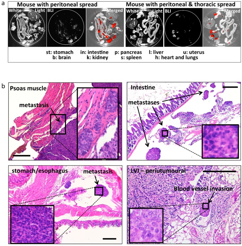
Figure 4.
Ex-vivo BLI and histologically confirmed metastases. (a) Representative images of post mortem ex-vivo BLI used to assess the metastatic spread to different peritoneal and extra peritoneal organs. Left: example of spread restricted to peritoneal organs. Right: example of spread to the heart/lungs (thoracic cavity). Strong BLI signal (white in the BLI images and red in the merged images) was associated with fat tissue adjacent to peritoneal organs (spleen, kidneys). (b) Representative images and histologic confirmation of tumour spread to the psoas muscle, intestine, stomach/oesophagus, LVI, and infiltrating tumour cells in the (anatomic location/organs are indicated in the images). Bar scale: 300 μm.

