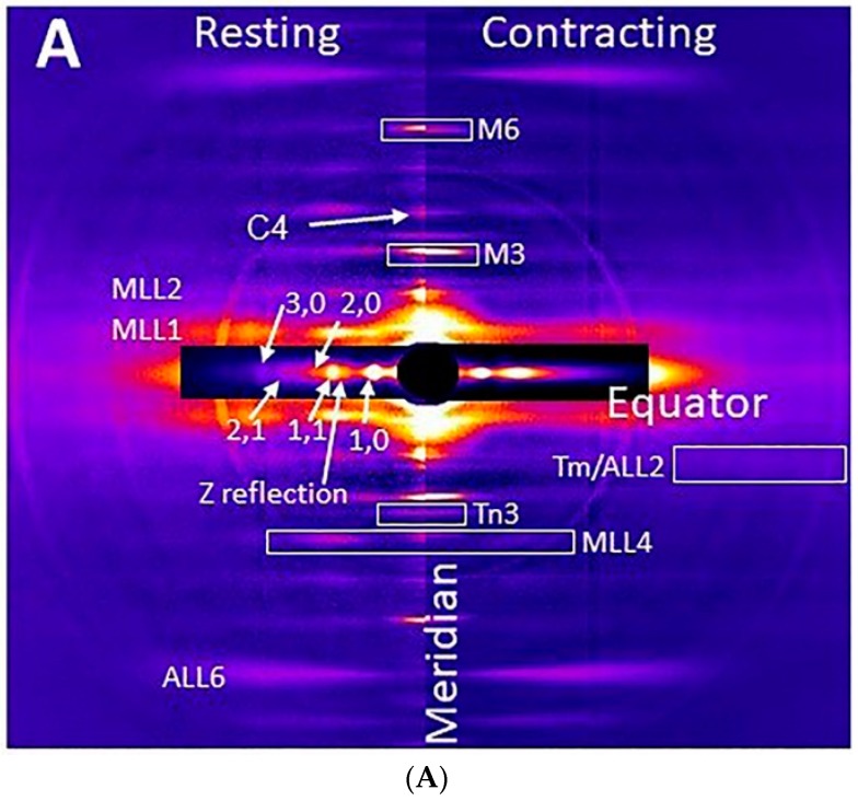
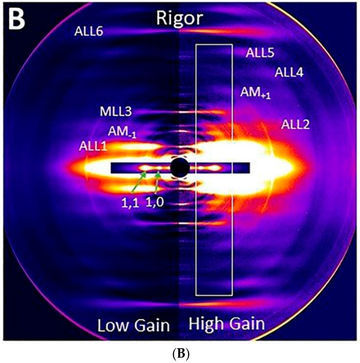
Figure 1.
X-ray diffraction patterns from mouse EDL muscle. (A) Patterns from resting (left) and contracting (right) mouse EDL muscle. The equatorial reflections and myosin layer lines (MLL) are as indicated. (B) X-ray pattern from muscle in rigor at low gain (left) and at high gain (increased 3-fold, right). The equatorial reflections and actin-based layer lines (ALL) are as indicated. The box indicates the range used for the integrated intensity trace shown in Figure 2. C4: 4th myosin binding protein C reflection; M3: third order myosin meridional X-ray reflection; M6: sixth order myosin meridional reflection. AM: actomyosin.


