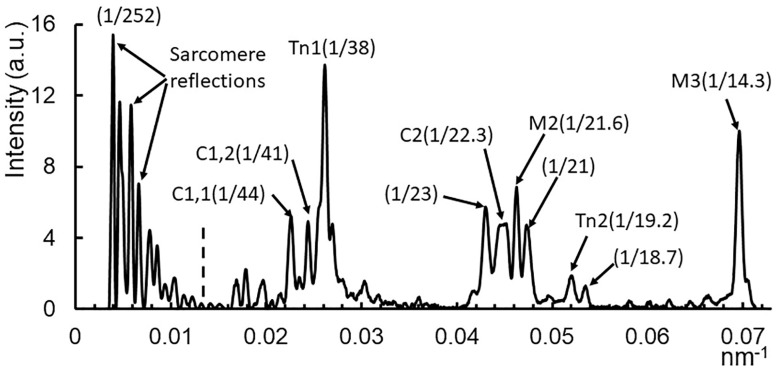
Figure 2.
Low order meridional reflections in resting muscle. The meridional reflections, including high order sarcomere repeat, M1 cluster, M2 cluster, and M3 reflection. Taken from resting muscle with a 9 m sample to detector distance. Integration region is from 0.03 nm−1 to 0.077 nm−1 in reciprocal space (white box in Figure 1B). Reflection intensities to the left of the dotted line are scaled down by a factor of 15 for visibility. C1: lowest angle myosin binding protein C reflection; C2: second myosin binding protein C reflection (doublet with C1); M2: second order myosin meridional X-ray reflection; M3: third order myosin meridional X-ray reflection; Tn1: 1st order troponin reflection; Tn2: 2nd order troponin reflection.

