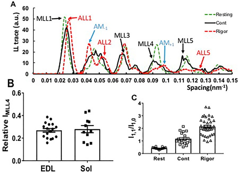
Figure 3.
Residual myosin layer line (MLL) intensity in X-ray patterns from contracting mouse muscle. (A) The layer line intensity traces from resting (green arrows), contracting (black arrows) and rigor patterns (red arrows) along the meridian integrated over a radial spacing range from 0.03 nm−1 to 0.077 nm−1. Both myosin (MLL) and actin (ALL) layer lines were present in contracting patterns, while all myosin layer lines were replaced by actin- or actomyosin-based layer lines in rigor patterns. (B) MLL4 remained at about 30% of its resting value in both EDL (0.27 ± 0.02, n = 17) and soleus muscle (0.28 ± 0.1, n = 11) during the plateau region of tetanic contraction. (C) Equatorial intensity ratio in resting (Rest), contracting (Cont) and rigor (Rigor) muscle.

