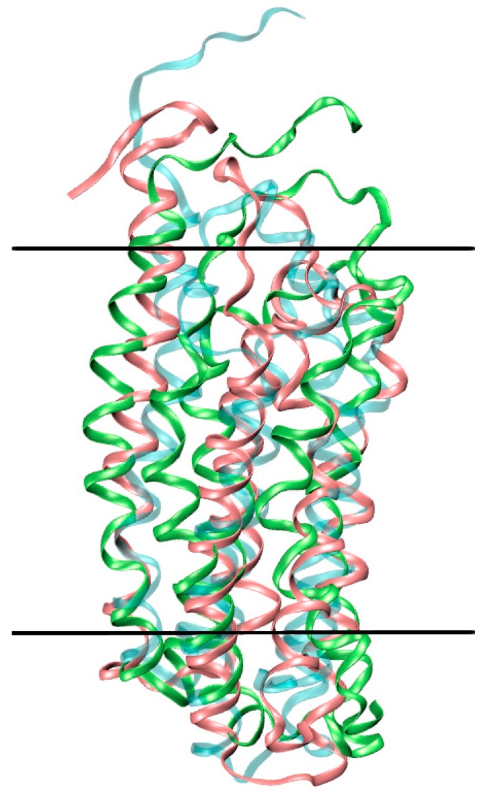
Figure 4.
Distortion of mTSPO_NMR_monomer in the lipid bilayer. The initial NMR structure (cyan) is superimposed to mTSPO_NMR_monomer in complex with PK11195 (green) and mTSPO_NMR_monomer without ligand (pink) after MD equilibration. The boundaries of the membrane are approximately shown as black lines.

