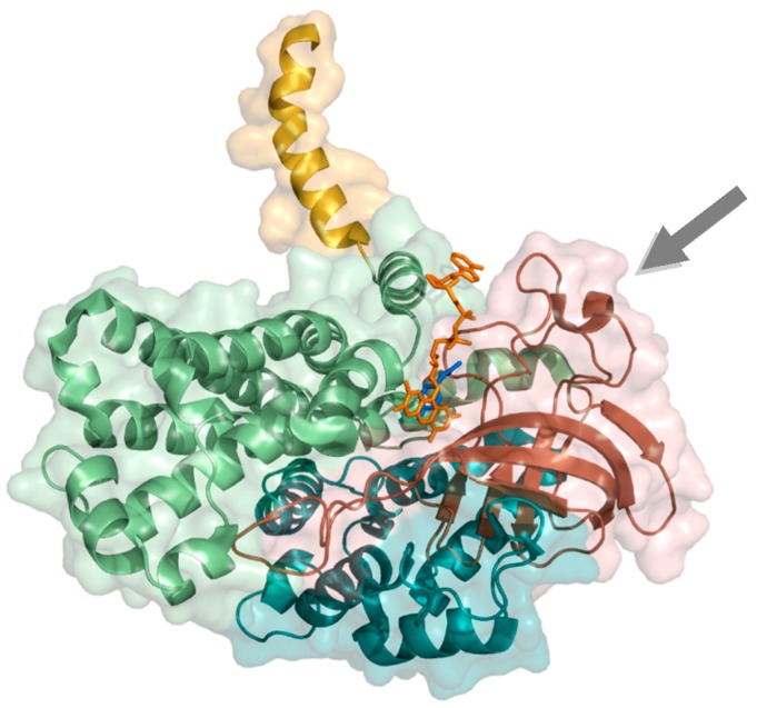
Figure 4.
Structure of 4-hydroxyphenylacetate 3-hydroxylase (HPAH) monomer (PDB ID: 2YYJ). Coloring is according to the N-terminal domain (teal), middle domain (brown), the C-terminal domain (pale green), and the C-terminal tail (yellow). The surface is indicated transparently, FAD (orange) and 4-hydroxyphenylacetate (blue). The loop region that allows recognition of the adenine moiety of FAD is indicated by an arrow.

