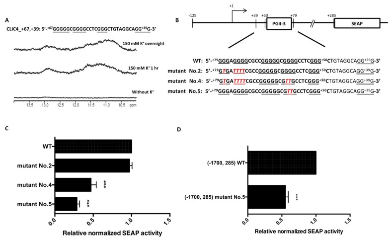
Figure 4.
Further disruption of the remaining possible G4 structure formed in the Mutant No. 2 reporter plasmid. (A) NMR analysis of CLIC4 +67 to +39, the possible G4-forming sequence in Mutant No. 2 in Tris-HCl buffer and 150 mM KCl, for 1 h and overnight; (B) Sequence of WT and mutants derived from Mutant No. 2—Mutants No. 4 and No. 5 contain the following G-tract at the 3′ end. The original PG4-3-forming region is shown in bold letters, G-tracts are underlined, and the mutated sequences in CLIC4 p(−125, +285) plasmids are marked in red italicized letters; (C) SEAP activity of CLIC4 p(−125, +285) mutants further disrupting the G4 forming motif in Mutant No. 2 were determined in A375 cells after transfection for 72 h. Data were expressed as the means ± SD of three replicates. *** p < 0.001 as compared to the WT. (D) SEAP activity of CLIC4 p(−1700, +285) mutant No. 5 in A375 cells after transfection for 72 h. Data were expressed as means ± SD of three replicates. *** p < 0.001 as compared to CLIC4 p(−1700, +285) WT.

