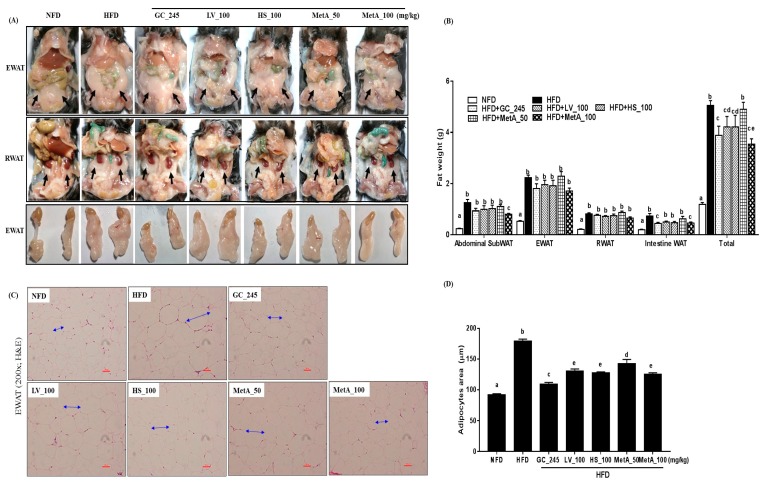
Figure 3.
Effect of LV, HS, and MetA on WAT weight and morphology in HFD-induced obese mice. Representative images (A), final WAT weight (B), hematoxylin and eosin (H and E) staining of WAT (C), and adipocytes area (D) in NFD and DIO mice after 9 weeks of 60% HFD feeding. Images were captured under a light microscope at ×100 magnification. Abdominal subWAT, abdominal subcutaneous fat; EWAT, epididymal white adipose tissue; RWAT, retroperitoneal white adipose tissue; Total, abdominal subWAT + EWAT + RWAT + intestine adipose tissue. HFD, 60% high-fat-diet control group; HFD + GC_245, HFD contains Garcinia cambogia extract (245 mg/kg)-fed group; HFD + LV_100, HFD contains lemon verbena extract (100 mg/kg)-fed group; HFD + HS_100, HFD contains Hibiscus flower extract (100 mg/kg)-fed group; HFD + MetA_50, HFD contains the MetA-mixed extract of lemon verbena plus Hibiscus flower (50 mg/kg)-fed group; HFD + MetA_100, HFD contains the MetA-mixed extract of lemon verbena plus Hibiscus flower (100 mg/kg)-fed group. Values are expressed as means ± SEM (n = 10). a–e Values within arrow with different letters are significantly different from each other at p < 0.05 as determined using Turkey’s multiple-range test.

