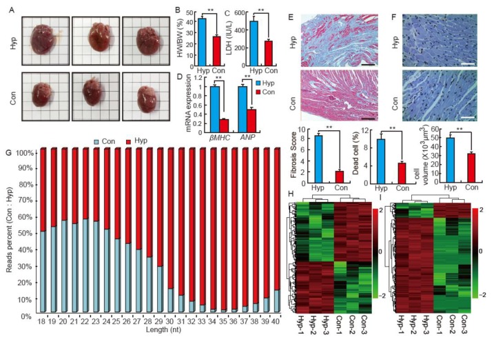
Figure 1.
Isoproterenol (ISO) induces cardiac hypertrophy and influences small RNA distribution in SD rats. (A) Images of whole hearts from control (Con) and isoproterenol-induced (Hyp) Sprague Dawley (SD) rats, n = 8; (B) The ratio of the heart weight to body weight (HW/BW) in Con and Hyp groups, n = 8. Data are means ± SD. ** p < 0.01; (C) The activity of serum lactate dehydrogenase (LDH) in Con and Hyp groups, n = 8; (D) Relative expression of mRNA of β-MHC and ANP. The qRT-PCR analysis was performed in triplicate with three independent samples; (E) Cardiac fibrosis evaluated by Masson trichrome staining. Scale bar = 200 μm, n = 8; (F) Histological analysis of heart tissue in Hyp and Con groups by Terminal deoxynucleotidyl transferase dUTP nick end labeling (TUNEL) staining. Scale bar = 50 μm, n = 8; (G) Reads distribution of small RNA (18–40 nts) in the Con and Hyp groups, n = 3; (H) Hierarchical clustering analysis for differentially expressed miRNA in Con and Hyp groups, n = 3; (I) Hierarchical clustering analysis for differentially expressed tRFs in Con and Hyp groups, n = 3.

