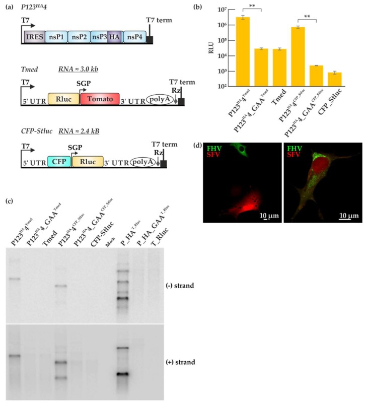
Figure 6.
Comparison of Semliki Forest virus (SFV) and FHV trans-replication systems. (a) Graphic representation of the SFV trans-replication system. In the two templates used here, Rluc is placed under the genomic or subgenomic promoter. The abbreviations and symbols are as in Figure 1. Non-structural proteins (nsPs) 1–4 represent the SFV replicase proteins. Tomato and cyan fluorescent proteins (CFP) are the fluorescent markers present on the templates. (b) BSR T7/5 cells were transfected with the SFV trans-replication systems and incubated at 37 °C for 16 h. Luciferase signals are shown as means from two independent experiments and the mock values have been subtracted. Error bars represent the standard deviation. ** designates p < 0.01 (Student’s t test). The templates used are indicated as superscripts together with the replicase plasmids. (c) Viral RNA synthesis was detected by Northern blotting using probes designed to detect negative and positive strands, using probes for the Rluc encoding region, common to all the systems. (d) BSR T7/5 cells were transfected with the two replication systems together (P123HA4+ Tmed + P_HA + T_eGFP) using Lipofectamine at 28 °C and RNA replication was detected by microscopy 40 h post-transfection. Green fluorescence represents FHV replication while red fluorescence represents SFV replication.

