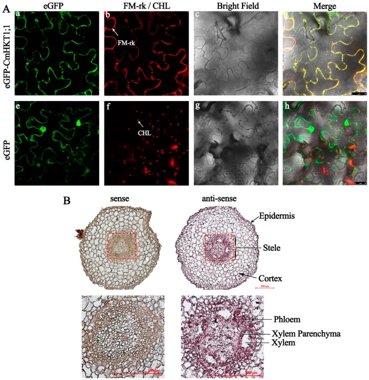
Figure 4.
Membrane and cell-type localization of CmHKT1;1. (A) Subcellular localization of eGFP-CmHKT1;1 in the pavement cells of N. benthamiana leaves. a, Green fluorescence from eGFP-CmHKT1;1. b, Red fluorescence from PM-rk-labeled membrane in eGFP-CmHKT1;1 expressing cells. c, Bright field image of the cell shown in a and b. d, Overlay image of a, b, and c. e, Green fluorescence from the free eGFP. f, Chlorophyll autofluorescence (CHL)-derived red fluorescence in eGFP-expressing cells. g, Bright field image of the cell shown in e and f. h, Overlay image of e, f, and g. (B) Localization of CmHKT1;1 mRNA in pumpkin root by in situ hybridization. Three true leaves aged pumpkin seedlings were subjected to 75 mM NaCl for 24 h before tissue collection. Cells in which transcript is present stain red-brown. Magnified views are shown below. Left panel, Sense probe. Right panel, antisense probe. The indication marks on the root cross section show the different tissues. Bars = 200 µm.

