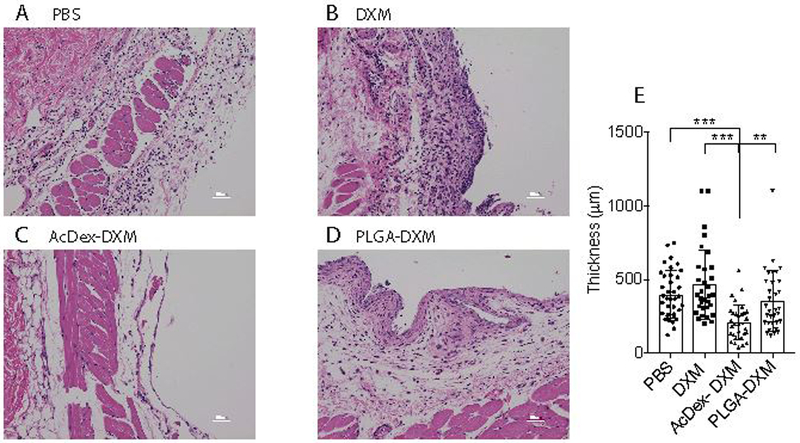
Figure 4. Reduced tissue infiltration after MSU crystals by AcDex-DXM as a 24-hour prophylaxis.

Mice were injected with prophylactic treatments (PBS control, DXM, AcDex-DXM or PLGA-DXM, 15 μg DXM/mouse) 24 hours before MSU crystal injections (3 mg in 0.5 ml PBS) into subcutaneous air pouches. After eight hours of stimulation, skin lining the pouch was preserved in 4% paraformaldehyde and subsequently stained by H&E. Representative images are shown in (A) MSU control, (B) DXM, (C) AcDex-DXM, and (D) PLGA-DXM. Scale bar represents 50 μm. The infiltrated cell area was quantified by Image J (E). The data is displayed as individual measurement points of 3 mice/group, with 6–16 sections per individual, and 32–37 distances per individual counted in total, ***p < 0.001, ** p < 0.01.
