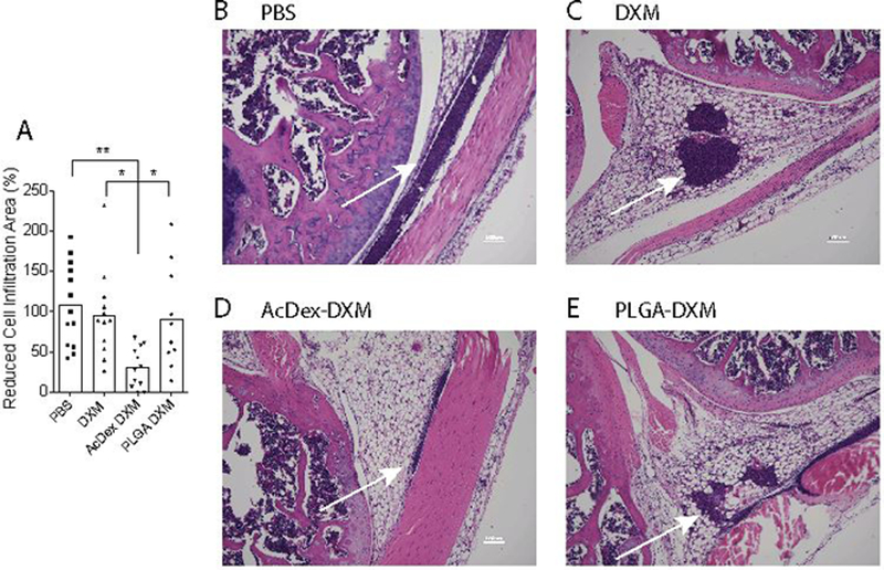
Figure 6. AcDex-DXM as an eight-day prophylaxis reduces inflammation in murine joints.

Mice were injected with prophylactic treatments (DXM, AcDex-DXM or PLGA-DXM, 10 μg DXM/mouse) eight days before MSU crystal injections (100 μg in 10 μl PBS) into murine knee joints. Eight hours after stimulation, knees were harvested, H&E stained and cell infiltration area was measured by Image J (A). (B-D) shows representative H&E stains of joints, scale bar is 100 μm. Arrows indicate cell infiltrations. In (A), dots represents individual data points pooled from two independent experiments (mean ± SEM), **p < 0.01,*p < 0.05.
