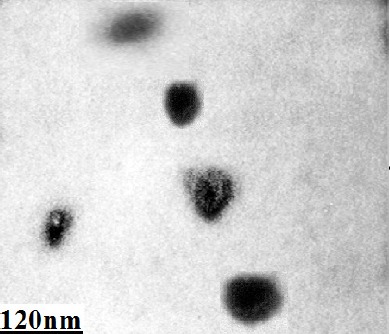
Figure 3.

Structural Analysis of MVs Derived from Bone Marrow-Derived Mesenchymal Stem Cells. MVs were isolated from CM of MSCs by differential centrifugation and observed by a transmission electron microscope (Philips Bio Twin, CM100, Netherlands) at 80 kV. MVs were seen as nano-sized vesicles in electron micrograph.
