
Fig. 2.
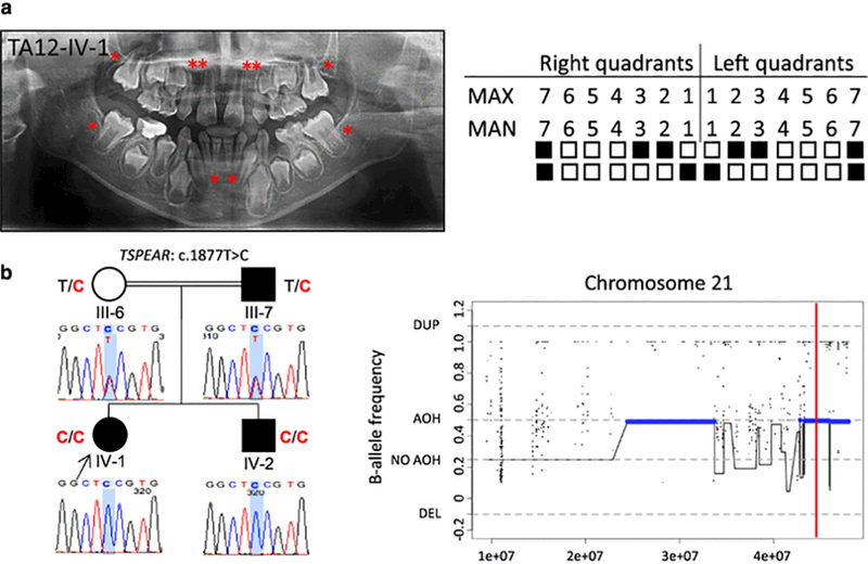
Clinical findings of TA12 family proband, segregation results, and AOH plots. (a) TA diagnosis was confirmed through clinical and radiographic examinations for patient TA12-IV-1. The panoramic radiograph of the proband was taken at 10 years 8 months. The missing permanent teeth are indicated as in Fig. 1a. (b) Pedigree and genotypes of family TA12. The same missense variant c.1877T>C (red font) in TSPEAR was identified in the proband (IV-1, arrow) and her affected brother (IV-2), while consanguineous parents (III-6 and III-7), an unaffected mother and affected father, harbor this variant in heterozygous state. This variant is located within one AOH region (~2.7 Mb). The total calculated AOH size in the proband is 231.807 Mb. Abbreviations: AOH, absence of heterozygosity; DEL, deletion; DUP, duplication.