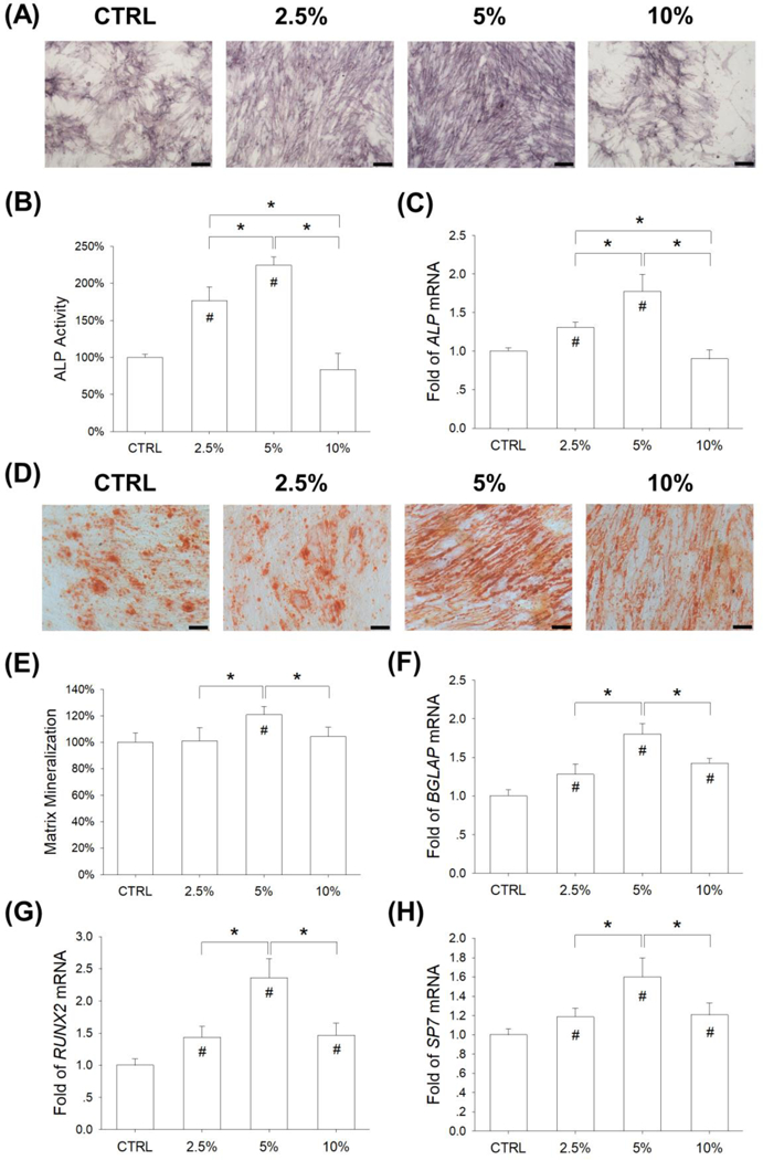
Figure 5.

Effects of mechanical stretch on the osteogenic differentiation BM-MSCs. Cells were exposed to cyclic stretch for 2 h per day at the magnitudes of 2.5%, 5%, and 10%. Cells cultured under static conditions served as the control (CTRL). (A) After a 7-day induction, intracellular alkaline phosphatase (ALP) stain was used as a marker of the early stage of osteogenesis. Scale bar = 200 μm. (B) Quantification of ALP activity in stretch-treated BM-MSCs. (C) The mRNA levels of ALP in stretch-treated BM-MSCs were quantified using RT-qPCR. (D) After 14 days of osteogenic differentiation, matrix mineralization was stained using Alizarin Red S. Scale bar = 200 μm. (E) Quantification of the stained mineral layers in stretch-treated BM-MSCs. (F-H) The mRNA levels of late-differentiated osteoblast marker genes, including BGLAP (F), RUNX2 (G), and SP7 (G) were quantified using RT-qPCR. Data are presented as the mean ± S.E.M. of four independent experiments (n = 4) in ALP activity, Alizarin Red S staining, and RT-qPCR experiments. Statistically significant differences are indicated by * where p < 0.05 between the indicated groups and # where p < 0.05 vs. the untreated cells.
