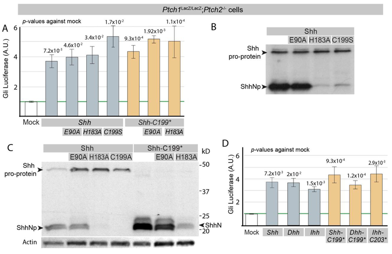
Figure 1. The Shh pro-protein can induce Gli-luciferase independent of Ptch1/2.
(A) Ptch1LacZ/LacZ;Ptch2−/− cells were transfected with Gli-Luciferase (Luc) alone (Mock) or cotransfected with Gli-Luc and Shh, Shh-E90A, Shh-H183A, Shh-C199S, Shh-C199*, Shh-C199*/E90A, or Shh-C199*/183A. Luciferase levels in mock transfected Ptch1LacZ/LacZ;Ptch2−/− cells were set at “1”. (B) Western blot analysis of Shh, Shh-E90A, Shh-H183A, Shh-C199S, all driven by the CMV promoter. (C) Western blot analysis of Shh, Shh-E90A, Shh-H183A, Shh-C199A Shh-C199*, Shh-C199*/E90A, and Shh-C199*/183A protein expression in Ptch1LacZ/LacZ;Ptch2−/− cells, using an antibody directed against the N-terminal domain of Shh. all driven by the CMV promoter, except Shh, which was driven by the EF1α promoter. (D) Ptch1LacZ/LacZ;Ptch2−/− cells were transfected with Gli-Luc alone (Mock) or co-transfected with Gli-Luc and Shh, Dhh, Ihh, Shh-C199*, Dhh-C199*, or Ihh-C203*. Luciferase levels in Mock transfected cells were normalized to 1. All error bars are s.e.m., p values (Student t-test, 2 tailed) against mock are indicated were relevant, n=4 (A), n≥3 (C), independent biological replicates, of triple or quadruple parallel experiments.

