
Figure 1.
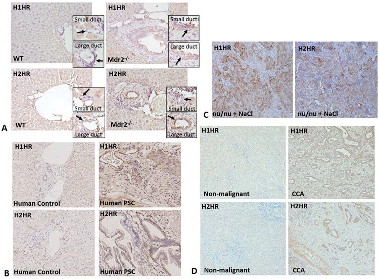
The expression of H1HR and H2HR in Mdr2−/− mice, nu/nu mice, and human PSC and CCA. Immunohistochemistry for H1HR and H2HR was performed in liver sections from WT, Mdr2−/− mice, nu/nu mice, human control and human PSC. H1HR expression was found in both small and large ducts of Mdr2−/− mice (but not in WT mice), with a more pronounced expression small (A). In WT mice, H2HR expression was low in large bile ducts, but absent in small bile ducts; however, in Mdr2−/− mice, H2HR expression was increased in large bile ducts, but not small (A). R Both H1 and H2 HR are expressed at low levels in human control liver sections, but the expression of H1 and H2 HR are increased in human PSC compared to controls (B). Tumors from nu/nu mice treated with saline expressed increased levels of H1HR and H2HR, particularly in CCA cells versus stromal cells (C). Both H1 and H2 HR are expressed at low levels in human non-malignant liver sections, but expression of both of these receptors is increased in human CCA when compared to non-malignant controls (D). Representative images are 20X magnification.