
Figure 5.
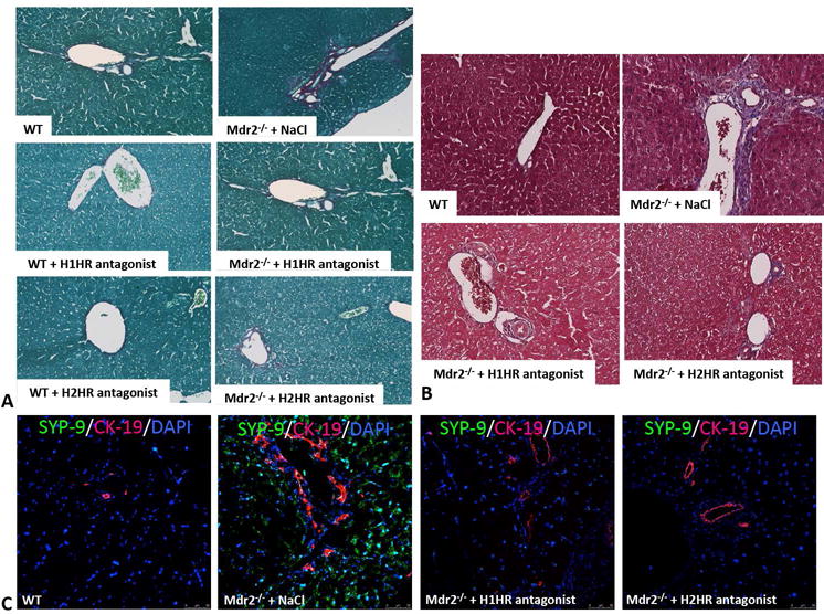
Assessment of hepatic fibrosis in WT and Mdr2−/− mice. In Mdr2−/− mice treated with saline there was increased collagen content and bridging fibrosis (as shown by Sirius Red and Masson’s Trichrome staining) when compared to WT mice; however, this was ameliorated in Mdr2−/− mice treated with either H1HR or H2HR antagonist when compared to saline treated mice (A and B). WT mice treated with H1HR or H2HR antagonists displayed little to no collagen deposition as shown by Sirius Red (A). HSC activation was measured by SYP-9 (green) immunofluorescence co-stained with CK-19 (red) to image bile ducts (C). There was an increase in activated HSCs in Mdr2−/− mice treated with saline when compared to WT mice, which was ablated in Mdr2−/− mice treated with either H1HR or H2HR antagonist when compared to saline treated mice (C). Representative images are 20X for colorimetric staining and 20X for immunofluorescence.