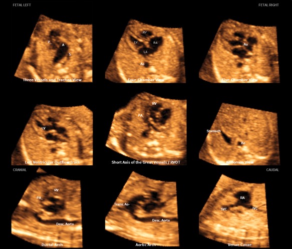
Figure 2.

Application of the FINE method to a fetus with tetralogy of Fallot at 23 weeks' gestation (diagnostic planes or VIS‐Assistance with automatic labeling are shown; also see Video 2). Six echocardiographic views were abnormal and demonstrate the typical features of this cardiac defect. The 3‐vessels and trachea view shows a narrow pulmonary artery caused by stenosis, while the transverse aortic arch is prominent. There is a “Y‐shaped” appearance of the great vessels. As is commonly noted in conotruncal abnormalities, the 4‐chamber view appeared normal in the diagnostic plane; however, VIS‐Assistance demonstrates a large ventricular septal defect (not shown here). The 5‐chamber view shows a ventricular septal defect. The left ventricular outflow tract view shows an overriding aorta, dilated aortic root, and ventricular septal defect. In the short‐axis view of great vessels/right ventricular outflow tract (obtained via VIS‐Assistance), the pulmonary artery is narrow with a tortuous ductus arteriosus. There is difficulty in visualizing a normal ductal arch. In the aortic arch view, the aortic root is dilated and there is a prominent ascending aorta. A indicates transverse aortic arch; Ao, aorta; Desc., descending; IVC, inferior vena cava; LA, left atrium; LV, left ventricle; P, pulmonary artery; PA, pulmonary artery; RA, right atrium; RV, right ventricle; RVOT, right ventricular outflow tract; S, superior vena cava; SVC, superior vena cava; and Trans., transverse.
