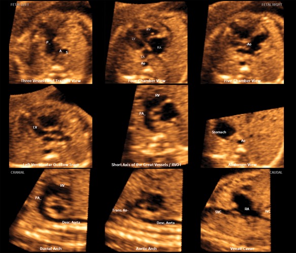
Figure 3.

Application of the FINE method to a fetus with coarctation of the aorta at 24 weeks' gestation (diagnostic planes or VIS‐Assistance with automatic labeling are shown; also see Video 3). Seven echocardiographic views are abnormal. The 3‐vessels and trachea view shows a narrow transverse aortic arch. In the 4‐chamber view, the left side of the heart is smaller than the right side; however, the left ventricle is apex forming. The right side of the heart appears enlarged, with the right ventricle being moderately dilated and hypertrophied. The 5‐chamber view shows similar findings to that of the 4‐chamber view. In addition, there is a narrow aortic root. The left ventricular outflow tract view shows a narrow aorta (obtained via VIS‐Assistance). In the short‐axis view of great vessels/right ventricular outflow tract, the cross‐section of the aorta is small compared to the pulmonary artery. The enlarged right atrium is apparent. The ductal arch view demonstrates that the cross‐section of the aorta is small compared to the pulmonary artery. In the aortic arch view, the coarctation is demonstrated as hypoplasia and narrowing of the transverse aortic arch as well as in the isthmus region. A indicates transverse aortic arch; Ao, aorta; Desc., descending; IVC, inferior vena cava; LA, left atrium; LV, left ventricle; P, pulmonary artery; PA, pulmonary artery; RA, right atrium; RV, right ventricle; RVOT, right ventricular outflow tract; S, superior vena cava; SVC, superior vena cava; and Trans., transverse.
