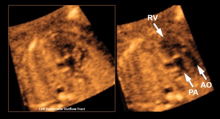
Figure 4.

VIS‐Assistance of the left ventricular outflow tract view in a 19‐week fetus with hypoplastic left heart, double outlet right ventricle, transposition of the great vessels, and fetal stomach on the right side (also see Video 4). The abnormal diagnostic plane (left panel) demonstrates a single vessel arising from the right ventricle that is consistent with the pulmonary artery due to its bifurcation. A second vessel could not be clearly identified. However, when VIS‐Assistance is activated (right panel), automatic navigational movements now demonstrate a second vessel (aorta) that is rightward and anterior and exiting the right ventricle. These 2 vessels are parallel and side by side, consistent with transposition. AO indicates aorta; PA, pulmonary artery; and RV, right ventricle.
