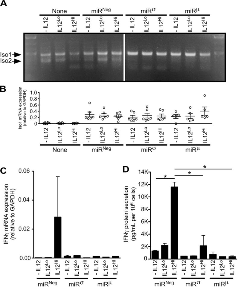
FIGURE 6.

Human IL12RB1 Isoform 2 positively regulates IL12-dependent IFNγ expression. Jurkat T cells were transduced via lentivirus with one of two microRNAs (miRσ and miRμ) designed to knockdown IL12RB1 Isoform 2 expression, without affecting IL12RB1 Isoform 1. The design and validation studies of miRσ and miRμ are shown in FIG S1; the lentiviral vector used for miR transduction also encoded GFP. Control Jurkat T cells were either left non-transduced (none), or transduced with a microRNA that does not target any human gene (miRNeg). GFP+ transductants were sorted and cultured in the presence of PHA and 3 different IL12 concentrations (-IL12, IL12Lo and IL12Hi). The cells in each culture condition were collected 2 days later, counted and used for (A-C) mRNA expression studies; cell supernatants were collected at the same time and used for (D) IFNγ ELISA measurements. (A) Multiplex amplification of Isoform 1 (Iso1) and Isoform 2 (Iso2) mRNA from stimulated miR-transductants. Shown is an agarose gel image that was used to visually confirm the knockdown of Iso2 in miRσ- and miRμ -transduced cells, relative to both non- or miRNeg-transduced cells (B) qRT-PCR measurement of Isoform 1 mRNA expression, as normalized to the GAPDH expression in the same cell preparations. Each open circle represents the data from an individual experimental replicate (6 experimental replicates per condition). (C) qRT-PCR measurement of IFNγ mRNA expression, as normalized to the GAPDH expression in the same cell preparations. Shown are combined data from all 6 experimental replicates. (D) IFNγ levels in the supernatants of stimulated miR-transductants, as normalized to cell count. Shown are combined data from all 6 experimental replicates; *, p<0.05 as determined by ANOVA analysis.
