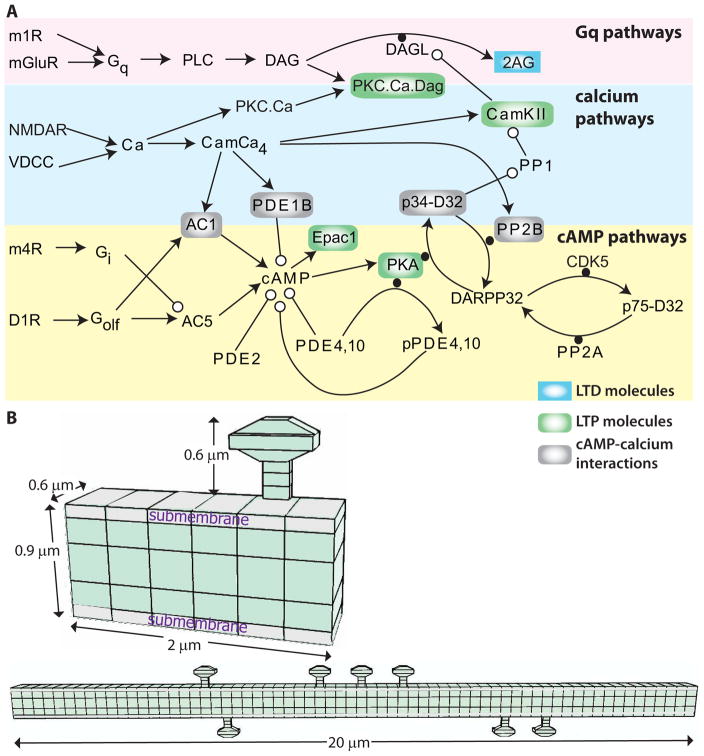
Figure 1.
Signaling pathways underlying synaptic plasticity in striatal direct pathway spiny projection neuron. (A) Dopamine D1 receptors (D1R) are coupled to the stimulatory subtype of GTP binding protein (GolfGTP), and lead to production of cAMP via adenylyl cyclase (AC). cAMP activates Exchange Protein Activated by cAMP (Epac) and protein kinase A (PKA). Both metabotropic glutamate (mGluR1/5) receptors and acetylcholine muscarinic type 1 (m1) receptors are coupled to Gq subtype of GTP binding protein (GqGTP) which activates phospholipase C (PLC), which produes of diacylgycerol (DAG), which is a substrate for diacylclyercol lipase (Dgl) and also activates calcium bound protein Kinase C (PKC). Acetylcholine also activates muscarinic type 4 (m4) receptors, which are coupled to inhibitory subtype of GTP binding protein (GiGTP) and inhibit the production of cAMP by adenylyl cyclase. Calcium (Ca) binds to various buffers including calmodulin which can activate protein phosphatase 2B (calcineurin; PP2B), phosphodisterase 1B (PDE1B) to degrade cAMP, and calcium calmodulin dependent protein kinase type 2 (CamKII). There are several interactions between these pathways: CamKII phosphorylates and inactivates diacylclyercol lipase, which produces the endocannbinoid 2 arachidonylgycerol (2AG). PKA phosphorylation of DARPP32 inhibits the protein phosphatase type 1 (PP1) that dephosphorylates CamKII. PLC: phospholipase C, PIP2: phosphoinositol bis-phosphate, PP2A: protein phosphatase 2A. (B) Morphology used for simulations, visualized using NeuroRDViz.py (https://www.gihub.com/neuroRD/NeuroRDviz). Most results used the 2 μm dendrite with single spine; simulations on spatial specifity used the 20 μm long dendrite with 10 spines (some attached to the same voxel). In both morphologies, the submembrane voxels are 0.1 μm high, the next layer of voxels are 0.2 μm high, and the central core voxels are 0.3 μm high. Diffusion is axial in the spines and two dimensional in the dendrite.

