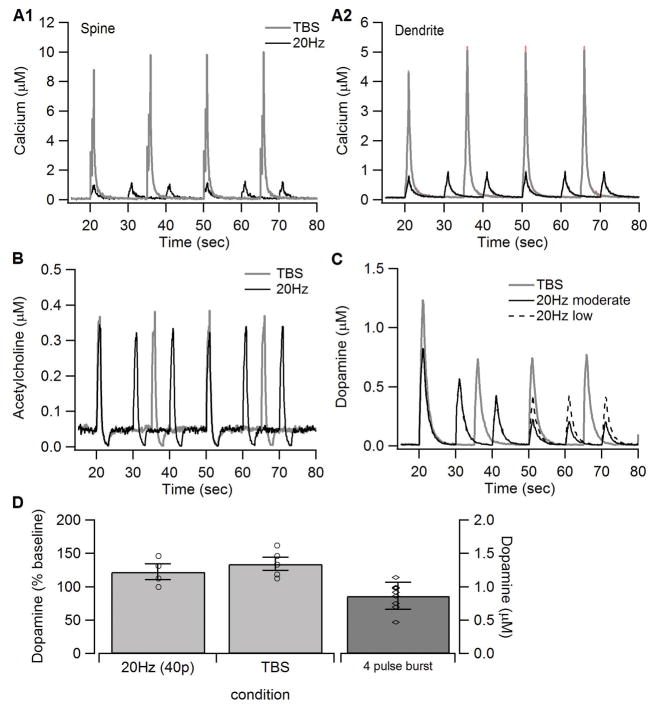
Figure 2.
Synaptic inputs to the model. The amount of calcium influx (A) was adjusted to reproduce the calcium concentration simuated using a biophysical model of a spiny projection neuron (Jedrzejewska-Szmek et al 2016). The concentration is considerably higher in the spine than in the dendrite, as observed experimentally. The concentration is higher for TBS than for 20Hz because the lower frequency stimulation depolarizes the neuron less than TBS. The acetylcholine profile (B) exhibited the burst and pause pattern observed in vivo. (C) Dopamine concentration used as input to the model. The reduction in dopamine for subsequent trains was based on measures of dopamine recovery kinetics (Mamaligas et al 2016), which shows that recovery of dopamine release is less after 10s intervals (used for 20Hz) than after 15s intervals (used for TBS). (D). Voltammetric measurements of dopamine used to constrain model dopamine input. A single burst of 4 pulses produces ~0.8 μM of dopamine. Giving 40 pulses of either TBS or 20Hz increases the dopamine transient by ~30% compared to the 4 pulse burst. Based on this data, dopamine input (shown in C) for TBS had a concentration of 1.2 μM. Since a train of 20Hz uses only 20 pulses, the dopamine transient for 20Hz was lowered compared to TBS.

