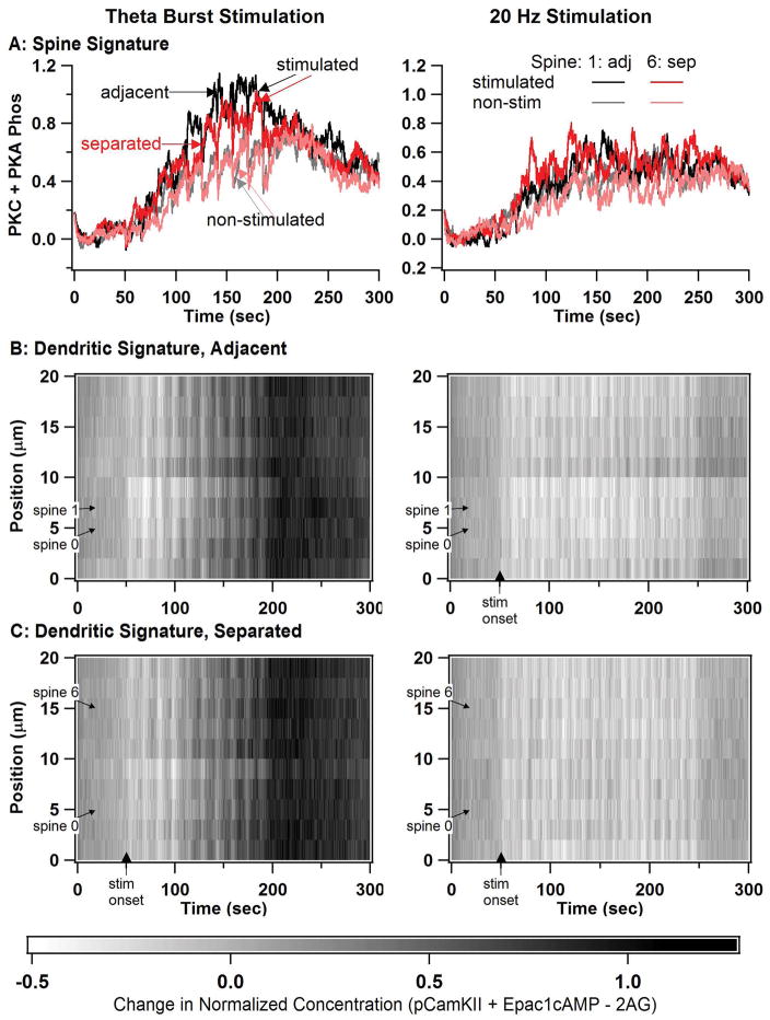
Figure 8.
Simulations in a long dendrite with multiple spines reveals spine spatial specificity for TBS but not for 20 Hz. In all simulations, spine 0 and either spine 1 or spine 6 are stimulated. (A) Spine signature (normalized sum of PKC activity and PKA phosphorylated proteins) reveal spatial specificity for theta burst stimulation, but not 20 Hz stimulation. Spine 1 is stimulated for the adjacent (adj) case and spine 6 is stimulated for the separated (sep) case. Only plots of spine 1 (black and gray traces) and spine 6 (red and pink traces) are shown, with the dark traces indicating the stimulated condition, and the lighter traces indicating the non-stimulated condition. (B) Dendritic signature differs between TBS and 20 Hz, but does not exhibit spatial specificity. Dendritic signature is calculated as normalized phosphoCamKII plus Epac1 minus 2AG. B1: two adjacent spines stimulated, B2: two separated spines stimulated.

