Figure 2.
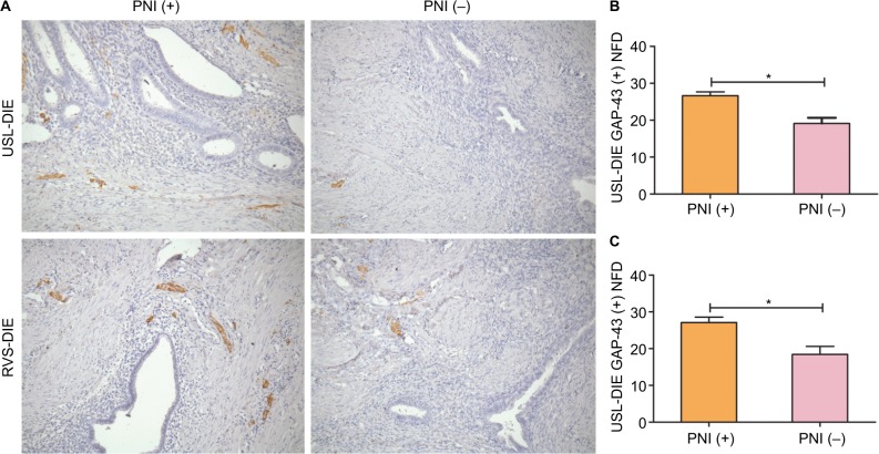
Detection of newly formed nerve fibers in lesions of USL-DIE and RVS-DIE.
Notes: Anti-GAP-43 was used to identify newly formed nerve fbers through immunohistochemistry (200× magnifcation). Newly formed nerve fibers were positively stained in USL-DIE and RVS-DIE lesions (A). NFDs of GAP-43 positively stained nerve fibers (shown in brown color) were significantly increased in lesions of PNI (+) group – (B) USL-DIE, P=0.001; (C) RVS-DIE, P=0.007 – compared with PNI (−) group. *P<0.05.
Abbreviations: USL-DIE, endometriosis infiltrating the uterosacral ligament; RVS-DIE, endometriosis involving the rectovaginal septum; Anti-GAP-43, antibody against growth-associated protein 43; NFD, nerve fiber density; PNI, perineural invasion.

