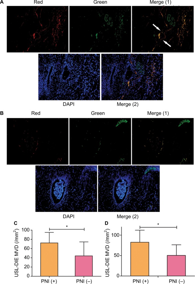
Figure 3.
Distribution of microvessels in endometriotic lesions with/without PNI.
Notes: Double immunofluorescence was performed to present microvessels and nerve fibers simultaneously. Microvessels were identified by antibody against CD31 (a specific marker of endothelial cells), which was red-stained, whereas nerve fibers were identified by antibody against PGP 9.5, which was green-stained. Nucleus was blue-stained with DAPI. More microvessels were found in endometriotic lesions of the PNI (+) group (A), compared to that of the PNI (−) group (B). Exact quantization of the MVD was acquired in both USL-DIE and RVS-DIE lesions. Further comparison analysis showed similar results: there was an increased MVD in endometriotic lesions of the PNI (+) group than that of the PNI (−) group (C and D).
Abbreviations: PNI, perineural invasion; MVD, microvessel density; USL-DIE, endometriosis infiltrating the uterosacral ligament; RVS-DIE, endometriosis involving the rectovaginal septum.

