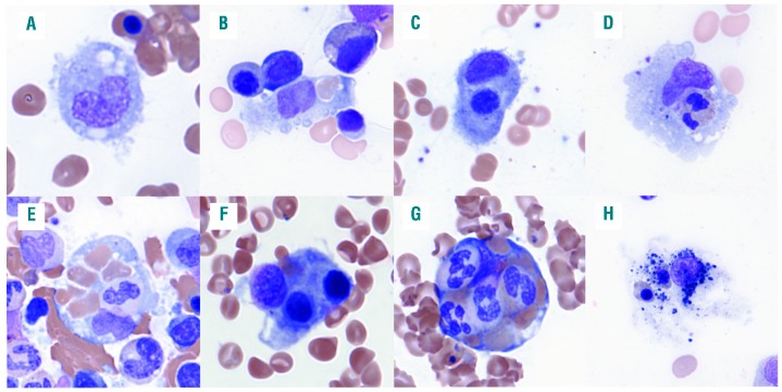
Figure 1.
Examples of hemophagocytosis in patients with hemophagocytic lymphohistiocytosis (HLH). (A) Histiocytes in patients with HLH often display rounded contour with cytoplasmic projections. (B-D) Hemophagocytes with a single ingested mature red blood cell (RBC), nucleated RBC progenitor, and granulocyte, respectively. Hematopoietic progenitor cells (HPCs) often contain single nucleated hematopoietic cells in addition to multiple mature RBCs (E); however, the presence of multiple nucleated cells within the cytoplasm of a single HPC (F and G) is highly predictive of the diagnosis of HLH. (H) An example of a histiocyte with degenerating nuclear debris, indistinct cytoplasmic contour, and equivocal intracytoplasmic nucleated RBCs that we do not consider to be a definite hemophagocyte.

