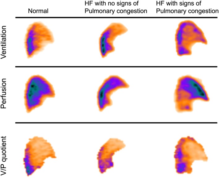
Figure 1.

Sagittal ventilation/perfusion (V/P) single‐photon emission computed tomography slices from the left lung of patients with heart failure (HF), with and without signs of pulmonary congestion, compared with a representative normal lung. In the normal lung and in the patient with HF but no signs of pulmonary congestion, pulmonary perfusion is predominantly distributed to posterior, that is, dependent parts of the lungs. In the lungs of the patient with pulmonary congestion and elevated pulmonary wedge pressure, perfusion is redistributed to anterior, non‐dependent, parts of the lung. Ventilation and V/P quotient images are included for reference purposes.
