Figure 1.

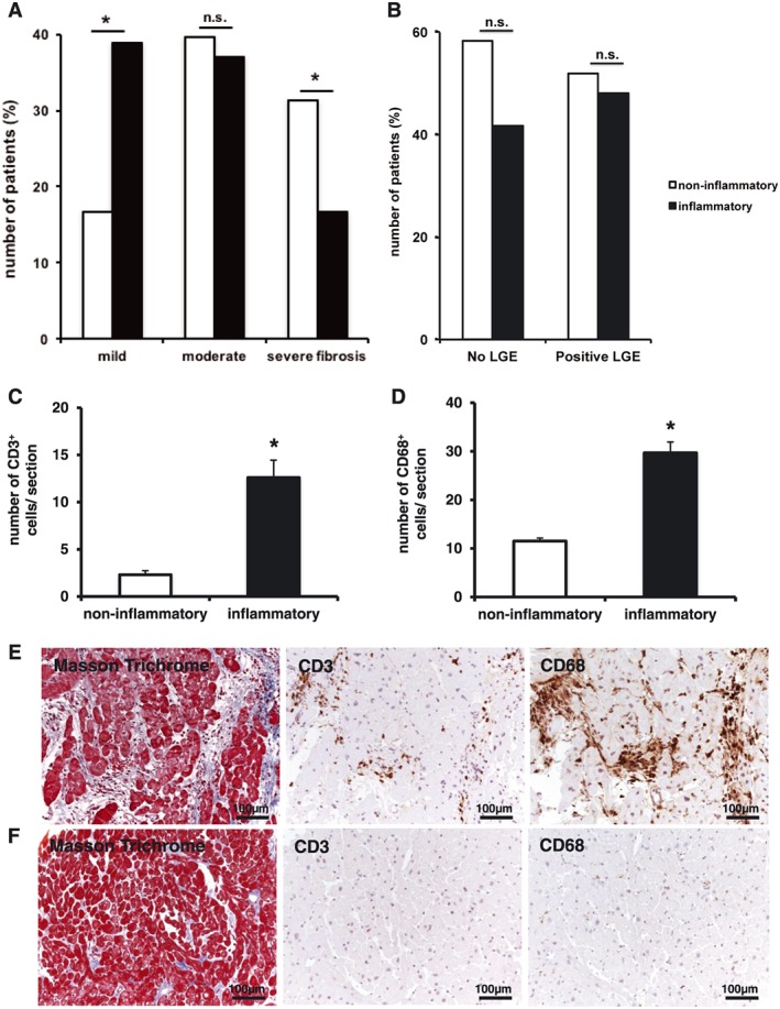
The degree of cardiac fibrosis and inflammation detected by endomyocardial biopsy is different in patients with inflammatory and non‐inflammatory cardiomyopathy, while the number of CD3‐positive T cells and CD68‐positive macrophages is increased in patients with inflammatory cardiomyopathy. According to the amount of fibrosis given in percentage (%) of fibrosis in relation to the total area of the biopsy, patients were categorized using tertile distribution defined as mild (Grade 1, 0–10%), moderate (Grade 2, 11–20%), and severe fibrosis (Grade 3, >20%). The number of CD3‐positive and CD68‐positive cells are given in cell number per section. Values are mean ± standard error of the mean; *P < 0.05. (A) Mild myocardial fibrosis was found significantly more often in patients with inflammatory cardiomyopathy (38.9% in inflammatory cardiomyopathy vs. 16.7% in non‐inflammatory cardiomyopathy, P = 0.024), while severe fibrosis was increased among patients with non‐inflammatory cardiomyopathy (16.7% in inflammatory cardiomyopathy vs. 31.3 in non‐inflammatory cardiomyopathy, P = 0.046). There was no difference in moderate fibrosis between 37% in inflammatory cardiomyopathy vs. 39.6% in non‐inflammatory cardiomyopathy, P = 0.675. (B) Positive late gadolinium enhancement (LGE) detected in cardiac MRI was present in 46 of the 102 patients (45.1%), but there was no difference between inflammatory and non‐inflammatory cardiomyopathy (P = 0.687). (C) The number of CD3‐positive T cells was significantly increased in patients with inflammatory cardiomyopathy compared with non‐inflammatory cardiomyopathy (12.6 ± 1.84 in inflammatory cardiomyopathy vs. 2.3 ± 0.43 in non‐inflammatory cardiomyopathy, P < 0.001). (D) CD68‐positive cells/macrophages were significantly increased in patients with inflammatory cardiomyopathy compared with non‐inflammatory cardiomyopathy (29.7 ± 2.23 in inflammatory cardiomyopathy vs. 11.5 ± 0.65 in non‐inflammatory cardiomyopathy, P < 0.001). (E) Representative myocardial tissue sections depict myocardial architecture in histological Masson's trichrome staining and positive expression of CD3 and CD68 on cells residing in the myocardium in inflammatory cardiomyopathy. (F) Representative myocardial tissue sections depict myocardial architecture in histological Masson's trichrome staining and positive expression of CD3 and CD68 on cells residing in the myocardium in non‐inflammatory cardiomyopathy.
